Kidney Stones and RIRS
Kidney stones and RIRS
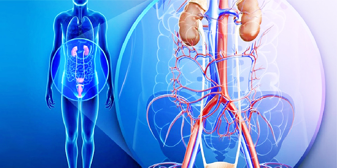
What Are kidney Stones?
Kidney stones, or renal calculi, are solid masses made of crystals. Stones can develop anywhere along your urinary tract, which consists of these parts
- Kidneys
- Ureters
- Bladder
- Urethra
Kidney stones are one of the most painful medical conditions. The causes of kidney stones vary according to the type of stone.
Types Of kidney Stones
Calcium oxalate
Calcium oxalate stones are the most common in India. High-oxalate foods include:
- potato chips
- peanuts
- chocolate
- beets
- spinach
However, even though some kidney stones are made of calcium, getting enough calcium in your diet can prevent stones from forming.
Uric acid
They can occur in people with gout or those going through chemotherapy. A diet rich in purines can increase chances of uric acid stones. Purine is a substance in animal proteins, such as fish, shellfish, and meats.
Struvite
This type of stone is found mostly in women with urinary tract infections (UTIs). Treating an underlying infection can prevent the development of struvite stones.
Cystine
Very rare. They occur in people with genetic disorder cystinuria.RISK FACTORS FOR KIDNEY STONES
Remember Mnemonic ‘BAD FOOODS’
-B: Bowel Diseases
-A: Animal proteins
-D: Dehydration (Consuming less water)
-F: Familial
-O: Obesity
-O: oxalate rich foods: potato chips, spinach,nuts, beet,tea
-O: Obstruction in urinary system
-D: Drugs (Diuretics, Antiseizure medicines, Calcium based Antacids
-S: Salt rich food ( Fast food, French fries, chips)
DIETERY ADVICE TO REDUCE STONE FORMATION
- Increase water intake to produce at least 2 liters of urine a day. Clear urine indicates sufficient hydration. This advice is not applicable to people with kidney failure, those with heart disease where water intake is restricted!
- Eat less salt and restrict diets rich in salt like fast food, French fries, potato chips
- One should NOT restrict intake of dairy products in history of calcium stones. Two to three servings in a day are acceptable.
- Limit the intake of animal proteins
- Limit intake of oxalate rich diet like spinach, nuts, chocolates, potato chips, peanuts, beet, colas, tea
- Tomatoes, as widely believed, are not a major risk factor for stones!
Calcium supplement: Dietary calcium in recommended doses does not increase risk of calcium stones. Restricting dietary calcium may in fact increase chances of stone formation. However your doctor may change the calcium salt to calcium citrate which is comparatively safer.
SYMPTOMS OF STONES
- Asymptomatic: Small stones in kidneys or one located in some locations of the kidney may be painless
- Pain: Sudden severe pain starting from back and radiating to front of abdomen or flank or genital area.
- Nausea or vomiting or
- Frequent urge to pass urine or
- Blood or brown colored urine.
- In case of associated urine infection there may be fever with or without rigors
DIAGNOSIS
- CT SCAN: Non Contrast CT scan (NCCT KUB) is the investigation of choice if available. Not to be done in pregnancy though.
- Ultrasound: Is an acceptable test if CT scan not available or in pregnancy. This may however miss stones in the ureter (Tubes draining urine from kidney to bladder).
- Removal of stones from kidney
- Treating tumors located inside the kidney. Urothelial tumors located inside the kidney can be removed/biopsied by RIRS
- Negligible blood loss
- Less pain as there is no cut or opening made
- Minimal injury to kidney as no cut or opening made in the kidney
- Safe to use in patients where open or conventional surgery is difficult or demanding like
- CT scan (CT KUB) to confirm location, size and number of stones and see anatomy of kidney and its drainage system
- Routine blood and urine tests
- Other tests as required by anesthetist to optimize the condition for giving anesthesia
- Fasting required for 4 to 6 hours before anesthesia
- Admission usually done on the day of surgery. Most discharged the next day, some can be discharged even the same day
- Fasting required for 4 to 6 hours
- Antibiotic given at the time of anesthesia
- Done under general anesthesia or spinal anesthesia.
- An endoscope (flexible ureteroscope) introduced through the urinary passage which provides a magnified view of inside the urinary system. The scope is advanced through the ureter to inside the kidney
- The stones are focused.
- A thin laser fiber is passed through the scope and advanced till the stone and laser energy focused over the stone reducing it into dust or small fragments.
- Some fragments retrieved using small specialized baskets passed through the scope.
- A thin tube called DJ stent left inside the kidney which extends from kidney to the urinary bladder. The purpose of stent is to allow any fragments or dust to flush out and to allow for healing.
- The duration of procedure will depend on the stone bulk
- The stent is usually removed after a gap of 1 to 4 weeks. The procedure is done by passing a scope inside the urinary passage (cystoscopy), which can be done under local or sometimes general anesthesia. During stent removal the surgeon may chose to have a look inside again to remove any residual fragments.
- A urinary catheter and bag is placed which is removed generally the next day if no fever.
- Oral intake of fluids allowed generally after 4 to 6 hours followed by a solid diet
- You are allowed to sit in the bed once effect of anesthesia is gone
- The urinary drainage tube (catheter) and bag are generally removed the next morning. Sometimes you can be discharged the same day if stone bulk is small and surgery not prolonged.
- You are allowed to take a bath next day and allowed walking or routine light work
- Because of the stent inside, expect following symptoms:
Other tests done in recurrent stone formers:
-Serum Uric acid, calcium, phosphorus
-Intact Parathormone(PTH) levels in blood
-24 hour urine for calcium,oxalate and citrate
-Urine routine and PH
TREATMENT
For pain:
-Painkillers of NSAIDS group (Ibuprofen,Diclofenac) : orally or in injection form.(Under supervision). Avoid in case of history of kidney dysfunction where other medicines like Tramadol are used.
-Avoid taking excess fluid during periods of severe pain as it may aggravate the pain or nausea and vomiting.
Medicines which help in passage of stone:
Medicines don’t help passage of stones located within the kidney. However small stones in the ureter may sometimes pass with medicines of alpha blocker group (the ones used for prostate treatment, like tamsulosin). The chances of stone passage depend on :
-Size of stone: more chance if smaller than 5 mm
-Location: more chances if located lower down in the ureter
-Duration : less chances if stuck for more than 3 weeks
WHEN NOT TO WAIT FOR STONES TO PASS WITH MEDICINES?
-Stones stuck on both sides: may lead to complete shutdown of kidneys and lead to acute kidney failure
-Associated infection: a stent may be placed in kidney (Double J stent) to clear infection and drain the kidneys
-Big sized stone with unlikely chances of passing through the ureter
-Stone stuck for more than a month: will start harming function of the kidney
-Single kidney: A stone blocking the solitary kidney can lead to acute kidney failure.
ESWL(Extra Corporeal Shock Wave Lithotripsy)
A non surgical method of stone removal by focusing strong waves generated by a lithotripter onto the stone by means of Xray or Ultrasound and breaking it into smaller fragments and waiting for their spontaneous passage through urine.
Advantages:
It is a non surgical method
Disadvantages:
-stones must be visualized on X Ray. !0 % stones not visible on X ray.
-Stones located in lower ureter. Shock waves may not reach adequately in lower ureter as that area is comparatively covered by bones of pelvis.
-Can’t be used in people with bleeding tendency or pregnancy
-May require multiple sittings
-stone clearance rates are very low if located in inferior pole of kidney. The stones may break but may not pass easily from that location
RIRS(Retrograde Intra Renal Surgery):
Stone removal done through a thin endoscope passed through the urinary passage and negotiated into the kidney. The stone fragmented with laser and fragments removed with a small basket introduced through the scope. There is no cut or opening needed on the body. A stent is left in place for a few days for any dust orfragments to pass through and helps in recovery.
Advantages:
-Very safe as no cut is made on the body and the risk of bleeding is negligible.
-Recovery is very fast as there is no cut or hole in the kidney
-No blood loss
Diadvantages:
Not the best option in patients with very large stone bulk where PCNL might produce higher stone clearance rates.
Dr.Aman Gupta is one of the few surgeons in India with one of the highest experience with this technique. Successfully performed more than 1200 of RIRS and mentored surgeons for this procedure from India and Overseas.
URSL(Ureteroscopic Lithotripsy):
Stone removal from ureter(tube of the kidney) using a rigid endoscope. This is done through the urinary passage under anesthesia and no cut or new opening is made externally. A stent is left in place for healing and washing out of residual dust or fragments.
PCNL(Percutaneous Nephro- Lithotripsy)
Through a small hole made in the back through the skin into the kidney, an endoscope(nephroscope) introduced into kidney, stones broken and removed. A tube (DJ stent) is left inside the kidney and sometimes an external tube as well from the back which is generally removed the next day. This procedure is preferred in a large stone bulk.
WHAT ARE THE SIDE EFFECTS OF THESE PROCEDURES
All these procedures are generally safe in expert hands. Side effects are:
1. Urinary infection
2. Residual stones: sometimes while breaking the stone, some fragments may migrate into areas which might be inaccessible or are so small that they are difficult to even grasp. Most of these residual fragments are harmless and need not be chased. Significant residual fragments may require another procedure to remove the stone. Incidence of residual stones is highest in ESWL.
3. Recurrent stones: Remember that a person who forms stones once is always at a risk to form stones throughout their lifetimes. All the precautions or dietery advise reduces the chances of recurrent stone formation but does not make it zero.
4. Stent symptoms: A Double J stent is left in place after all the endoscopic procedures and some cases of ESWL with a large stone bulk. The stent causes some blood in urine (which is harmless), frequent urination and some discomfort in the back while passing urine. These symptoms go away after the stent is removed.
5. Bleeding: PCNL involves making a puncture in the kidney, so bleeding is a risk. Some may require blood transfusion or rarely the bleeding vessel needs to be clotted artificially by introducing an artificial clot/coil through a peripheral vein by an interventional radiologist. A person should avoid heavy physical activity for a period of 2 to 3 weeks after this procedure. Inform your surgeon if you are on any blood thinners (warfarin, aspirin, clopidogrel) painkillers, Vitamin E all of which can increase the chances of bleeding. Under supervision, these medicines have to be stopped 3 to 7 days before PCNL. Also inform if you have a history of bleeding disorders like hemophilia or advanced liver diseases.
In selected cases , RIRS may be a better procedure in such situations which carries a minimal risk of serious bleeding
6. Serious Injury to ureter or kidney occur uncommonly and may require conversion to open surgery or multiple other surgeries.
7. Failure to remove stone: This is one situation which most of the patients are concerned about. Why it happens. Explanation is as below: Sometimes the anatomy of a person is such that the instruments are unable to go into the kidney or ureter. Most of such situations are encountered only once a surgeon has actually had a look inside the urinary system and may not be diagnosed beforehand on scans. This can happen as high as one out of ten times! In such situations, it might not be safe to remove the stone in the same sitting, and the surgeon just leaves a stent behind in the kidney. The stent serves two purposes: it bypasses the blockage if any caused by the stone thereby preventing further harm to the kidney, and it passively opens up the ureter thereby making instrumentation easy after a few days. The procedure for stone removal is done in a second stage after a few days.
URINARY BLADDER STONES
Usually found in malnourished children or men with urinary tract obstruction
Symptoms
Poor or interrupted stream of urine
Blood in urine
Urine infection
Treatment
Cystolithotripsy:
Endoscopic removal of stone through urinary passage or through a small hole through the bladder made just above the pubis.
The underlying urinary obstruction if present is also treated simultaneously (like prostate or narrowing or stricture of urinary passage)
Dr.Aman Gupta has performed more than 2500 of Laser procedures for stone removal including more than 1000 RIRS which makes him one of the most experienced in this procedure in the country. He also has mentored a few urologists for RIRS from India and overseas
WHAT IS RIRS?
Retrograde Intra Renal Surgery is a technique for removal of stones from ureter and kidney without making any cut or hole on the body. It is done by a Urologist using a thin flexible video-endoscope(flexible ureteroscope)through the urinary passage, along with Holmium laser to dust the stones.
WHAT IS RIRS USED FOR?
WHAT ARE THE ADVANTAGES OF RIRS?
No cut or opening made on the body. It is done through urinary passage.
Faster recovery
-those with bleeding disorders like liver dysfunction, hemophilia
-Obese patients where positioning will be difficult for conventional open surgery or PCNL
HOW IS RIRS PERFORMED?
Preoperative Tests:
Operative procedure
Post operative advice
-some blood in urine when you pee
-frequent urge to pass urine
-some discomfort in the back or the urinary passage while passing urine.
Some oral antibiotics or painkillers are advised generally till the time stent is not removed
You are called again on a subsequent date for stent removal.
